"What lies behind us, and what lies before us are small matters
compared to what lies within us."
- Ralph Waldo Emerson
Monday, February 7, 2011
Interpreting Urinalysis Results
At one point in our jobs or careers we may have to be subjected to the slight humiliation of a urinalysis. But what do the results mean? Here are some definitions that may clear it up for you.
Glycosuria: having glucose in the urine
Levels of glucose in the urine should be minimal (less than 40mg/dl) or none at all. Glucose in urine could be caused by carbohydrate heavy meals, too much sugar, or could be an indicator of diabetes mellitus.
Bilirubinuria: presence of bile pigments and bilirubin in urine
This can be caused by erythrocyte (red blood cell) breakdown
(hemolytic anemia), blockage of the bile duct or liver damage from hepatitis or cirrhosis.
Ketosis: ketone bodies in urine
This can be caused by our bodies using up fat storage that is meant for times of starvation or in individuals with diabetes mellitus or in an abnormally high fat diet.
Hematuria: red blood cells have ruptured and are present
Instead of having erythrocyte destruction, as in bilirubinuria, the erythrocytes have ruptured such as during a menstrual cycle or kidney stones.
Albuminuria: presence of a common blood protein called albumin
This can be due to diabetes mellitus, renal damage (kidney disease), extreme physical activity or hypertension.
Pyuria: elevated levels of leukocytes
Leukocytes are white blood cells which are used to fight pathogens in the body. High levels of leukocytes could mean a urinary tract infection or bladder or kidney infection.
***These are my notes from physiology lab, and are by no means a way to diagnose yourself or others.
Friday, January 28, 2011
Microbiology: The First Golden Age
The Scientific Method
Observation
Hypothesis
Experiments to test Hypothesis
Interpretation of test results
Conclusion to prove or disprove the hypothesis
Anton van Leeuwenhoek (1632-1723)
First to see "animalcules" or protozoa in 1674
Reported existence of bacteria in 1674
Made his own microscopes for inspecting the cloth he bought and traded. He created the first microscope able to see microbes. Unfortunately he did not teach anyone his trade, so the method to make these microscopes died with him.
No link was made between "animalcules" and illness
Spontaneous Generation
Doctrine that lifeless substances give rise to living organisms (see Controversy over Spontaneous Generation
Scientists Redi, Needham, Spallanzani and Pasteur all dealt with this phenomena
First observations:
wheat bran + rags = mice
meat + time = maggots
Redi (1670)
Created experiment that proved that flies were required for the formation of maggots on meat by using lace to cover a jar of meat.
Needham (1748)
Used two cork flasks, one slightly heated, one not. He observed that after sitting for some time both still had microorganisms in them.
Problem: He did not heat the one enough to kill anything, and he did not know how many bacteria he started with in the first place.
Spallanzani (1767)
Used FOUR flasks with mutton boiled infusion.
One open to air
One corked
One heated slightly with cork
One boiled longer, sealed by melting the glass
He revealed that the 4th, boiled and sealed flask did not grow microorganisms.
Needham countered that because Spallanzani had sealed the flask he had deprived it of it's "life force" or oxygen so therefore of course it didn't grow anything.
Louis Pasteur (1870)
1. Disproved Spontaneous Generation with his neck flask experiment. Allowed air in but gravity prevented microorganisms from getting into the broth. Once tipped so that the broth touched the dip in the neck, microorganisms grew.
2. Thought of the Germ Theory of Disease
3. Proved that yeast makes wine with grapes
4. Developed a vaccine for rabies
5. Developed immunization techiniques
Robert Koch (1875) pronounced "coke"
1. Proved the Germ Theory of Disease
Created Koch's Postulates: (to prove a microbe is the cause of a disease)
1. Always observe organism in diseased animal
2. Isolate organism in pure culture
3. Inoculate healthy animal and re-create the disease
4. Re-isolate the organism from the experimentally infected animal
2. Developed Pure Culture Technique
Because of Hess, he adapted Agar, used by Hess to make jelly, as his solid culture medium.
Agar is a powder from seaweed; solid at room temp, and it will not be degraded by bacteria.
Finally practices were being used to reduce the transmission of infectious disease:
sterile practices in hospitals
pasteurization of dairy products
insect control
care in preparation of food
sanitation improvements
personal hygiene
Spinal Cord/Brain/Nervous System Facts
The four spinal nerve plexi and one nerve from each plexus are:
| Cervical - | Phrenic nerve |
| Brachial - | Radial nerve |
| Lumbar - | Femoral nerve |
| Sacral - | Sciatic nerve |
The lateral ventricles are connected to the third ventricle by the right and left interventricular foramina while the third ventricle is connected to the fourth ventricle by the cerebral aquaduct
The central sulcus divides the frontal/parietal lobes.
The longitudinal fissure divides left/right hemispheres of the cerebrum.
The parietiocciptal sulcus divides parietal/occipital lobes.
The lateral fissure divides the parietal/temporal lobes.
The transverse fissure divides parietal/occipital lobes.
Many spinal tracts undergo decussation to communicate across the mid-sagittal plane.
Rostral means towards the nose.
Spinal nerves are considered mixed nerves because they consist of bundles of axons carrying sensory information as well as bundles of axons carrying motor information.
The gray matter of the spinal cord is located in the deep layers of the spinal cord and deep to the white matter.
The medulla oblongata is the portion of the brainstem most involved in control of heart rate, blood pressure, and respiratory rate.
CSF (cerebrospinal fluid) is produced in areas known as choroid plexi.
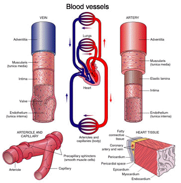
Blood Vessels
Capillaries - exchange vessels
Viens - blood to the heart
The vessel wall
Tunica interna
Simple squamous epithelium or endothelium
Loose connective tissue
Tunica media - muscle layer
Smooth muscle and collagen fibers, some elastic fibers in arteries
Provides vasoconstriction and vasodilation
Tunica externa - gives the vessel it's round shape
Loose connective tissues that anchors vessel in place
Resistance vessels - maintain shape when empty or cut
Conducting or elastic arteries - large
Tunica media has many elastic fibers
Distributing or muscular arteries
Distribute blood to specific organ
Mostly smooth muscle
Usually have individual names
Resistance vessels
Smallest are called arterioles
Some end in metarterioles or precapillary sphincters (gate)
Arterial Sense Organs
Monitor blood pressure and blood chemistry and transmit to the brainstem to regulate heartrate, vasomotion, and respiration.
Carotid sinuses - baroreceptors in carotid artery and innervated by glossopharyngeal nerve to help adjust heartrate and blood pressure (monitors bp)
Carotid bodies - chemoreceptors in carotid artery that monitor carbon dioxide, oxygen and pH levels of blood, send signal via vagus and glossopharyngeal nerves to adjust respiration and kidney function
Aortic bodies - same as chemoreceptors in carotid bodies, but located in the aortic arch
Capillaries
Exchange vessels
Consist of only tunica interna
In every tissue except: tendons, ligaments, cartilage, epithelia, and the cornea and lens of the eye
Types of capillaries: (based on cellular junctions and permeability)
Continuous capillaries - BBB (Blood Brain Barrier), stomach lining
Fenestrated capillaries - most common, skeletal muscles
Sinusoids - more holes than cells, in hypothalamus and liver
Capillary beds
Thoroughfare channels - capillaries to arterioles
Tendons and ligaments are avascular so there is little blood supply to give nutrients and allow them to heal. This is why it takes less time for bones to mend than sprains or strains.
Also, the crazy German scientist who created Body Worlds that travels around the globe used a process called Plastination to keep the bodies the way they are. This works at an intercellular level allowing only blood vessels or muscles for example to be kept in place while all the other bodily tissues go away.
VeinsCapacitance vessels - large capacity
Postcapillary venules - mostly tunica interna, somewhat permeable
Muscular veins - 1 to 2 layers of tunica media
Medium veins - have individual names and all tissue layers
Venous layers - many femoral valves (to prevent varicose veins)
Venous sinuses - normal varicose veins where blood is SUPPOSED to pool
Large veins
Mostly tunica externa
60% of our blood is in our veins @ all times because it is more difficult and takes longer for the blood to go against gravity back up to the heart from our limps.
Circulatory Routes
Typical route:
heart - arteries - capillaries - veins - heart
Exceptions to the typical route:
Portal systems
Anastomosis
Arteriovenule anastomosis - arteriole to venule
Arterial anastomosis - two arteries to one capillary bed
Venous anastomosis - more than one vein draining capillary beds
Blood Vessel Facts
The three major branches from the aortic arch are the right brachiocephalic trunk, left common carotid artery, and the left subclavian artery.The medial cubital vein is a connection of the radial, ulnar, basilic and brachial veins, is found on the anterior portion of the elbow and is a common site for blood draws.
The renal artery supplies the kidneys with blood while the hepatic artery supplies the liver with blood.
The hole in the diagram that major blood vessels pass through is the aortic hiatus.
The longest blood vessel in the body is the great saphenous vien.
The portion of the descending aorta which is superior to the diaphragm is the thoracic aorta.
Blood Flow Through the Heart
| 1. Systemic circuit 2. Vena cava (superior and inferior) 3. Right atrium 4. Tricuspid valve 5. Right ventricle 6. Pulmonary semilunar valve 7. Pulmonary arteries (deoxygenated) 8. Pulmonary circuit (lungs) | 9. Pulmonary veins (oxygenated) 10. Left atrium 11. Bicuspid (Mitral) valve 12. Left ventricle 13. Aortic semilunar valve 14. Aorta 15. Systemic circuit again |
|---|
The Heart!
There should always be 5-6 liters of blood in the human body
Gross anatomy
The heart wall consists of 3 different tissues:
Epicardium - the surface of the heart
Myocardium - contractile muscles of the heart
Cardiocytes
Endocardium - internal lining, simple squamous epithelium
Heart valves
Ensure one-way flow of blood
Consist of 2 or 3 cusps
Tricuspid valve - between right atrium and right ventricle
Bicuspid (Mitral) valve - between left atrium and left ventricle
Chordae tendineae - 'cords of tendon'
Papillary muscles - stabilize heart valves
Pulmonary semilunar valve - between systemic circuit and right atrium
Aortic semilunar valve - between left ventricle and aorta
75 bpm on average in a resting adult
Myogenic - muscle cell, generates electricity
Autorhythmic - creates electricity while ignoring brain
Conduction system consists of modified cardiocytes
Sinoatrial (SA) node - group of myogenic cells
Atrioventricular (AV) node
AV bundles
Left and right bundle branches
Moderator band of R. ventricle
Purkinje fibers - fingerlike projections into ventricles
Cardiac muscle
Striated, but short, thick and branched
Fibers contain only one centrally placed nucleus
Cardiocytes are joined end to end by intercalated discs - allows cells to communicate
Cranial Nerves
|
Cytology
The Cell Theory:
All organisms are composed of cells
Cells are smallest physiological unit
Organism activity is based on cellular activity
Cells come only from preexisting cells
All cells have similar molecular characteristics
Cell Shapes and Sizes:
Squamous - fried egg shape
Cuboidal
Columnar
Polygonal
Stellate - star shaped
Spheroid/ovoid
Discoid
Fusiform
Fibrous
Basal - bottom, 'basement'
Apical - top, surface
Cells measured in micrometers - RBC's are 7-8 micrometers
The Outer-Cellular Components:
ICF - intracellular fluid
ECF - extracellular fluid
The Plasma Membrane:
Holds material in and out of cell
Many physiological functions
Fluid mosaic model - varied, nothing is solid
Phospholipid bilayer - fat w/ a phosphate attached
Hydrophilic heads
Hydrophilic tails
Cholesterol - makes hormones
Glycolipids - combination of sugar and fat
Membrane Proteins:
Integral proteins - pass all the way through membrane
Peripheral proteins - only found on either side
Functions:
Receptors - allows chemicals in/out
Enzymes - proteins, speed up cells
Channels - passageway from ICF to ECF
Cell identity markers - peripheral in ECF
CAMS - cell adhesion molecules, keeps things in place
Carriers - integral proteins (specific channels)
Membrane transport - how material gets back and forth through membrane
Outside the Cell Membrane:
The Glycocalyx - slim layer, ID's cell, keeps in place
Glycolipids and glycolipids
Functions - enables movement
Cellular Extensions:
Microvilli - smallest
Cilia
Flagellum - largest
Intercellular junctions:
keeps cells together
Tight junctions - share a membrane (fence)
Desmosomes - peripheral, snap connection/velcro
Gap junctions - integral, direct communication
Cytoplasm:
Micro filaments
Intermediate filaments
Microtubules
The Nucleus - boss
Nuclear envelope and pores, chromatin, nucleoplasm,
Endoplasmic Reticulum - production facilities
Rough - produces proteins
Smooth - produces lipids and carbohydrates
Ribosomes - make protein out of amino acids
Free - mobile in ICF
Fixed - attached
Golgi Complex - packaging, addresses proteins
Lysosomes - gets rid of waste, 'stomach of the cell'
Peroxisomes - gets rid of H2O2 and alcohol specifically
Mitochondria - powerplant, convert to ATP
Crista (folds) - double membrane, inner, large surface area
Matrix - fluid in mitochondria
Centrioles - expansion devices, makes new cells (divides)
Inclusions - storage sheds after product is made
Fluid different in chemical composition than ECF (sodium)
Nervous Tissue
Overview of the Nervous System
* Body must communicate on a cellular level
o Nervous system and endocrine system
Functions of the Nervous System
* Receive information about changes inside and outside the body
* Process this information and determine appropriate response
* Issue commands to cells to carry out the response
Hierarchical Organization of the Nervous System
* Central Nervous System
o Brain
o Spinal cord
* Peripheral Nervous System
o Sensory (afferent) division
+ Visceral sensory division
+ Somatic sensory division
o Motor (efferent) division
+ Visceral motor division (autonomic nervous system)
# Sympathetic division
# Parasympathetic division
+ Somatic motor division
Neurons
* Functional unit of the nervous system
* Properties
o Excitability
o Conductivity
o Secretion of neurotransmitters
* Functional classes of neurons
o Sensory (afferent) neurons
o Interneurons (association neurons)
o Motor (efferent) neurons
Structure of a Typical Neuron
* Soma
o Organelles
o Neurofibrils
o Nissl bodies
o Inclusions (lipofuscin)
* Dendrites
* Axon
o Axon hillock
o Collaterals
o Axoplasm and axolemma
o Terminal arborization
+ Synpatic knobs
The Muscular System
There are 3 different types of muscles
Skeletal Muscle -
Functions of Muscle:
Movement - can contract and change
Stability - keeps the bones together
Communication - facial expressions (smile, speak)
Control of body openings - voluntary
Heat production - keep body 98.6 degrees, muscle contractions (shiver - skeletal muscle)
Properties of Muscle:
Excitability - can work with electrical signals
Conductivity - can conduct electricity
Contractibility - ability to shorten
Extensibility - some ability to stretch
Elasticity - ability to return to original state
General Anatomy of Skeletal Muscles:
Made of muscle fibers, connective tissue, nervous tissue, and blood vessels.
Connective tissues:
Endomysium - single layer collagen that surrounds the inside like a cell wall
Perimysium and fascicles - end of each muscle cell, divided by perimysium
Epimysium - holds all the fascicles together in a muscle
Deep fascia
Superficial fascia - hypodermis
Fascicles and Muscle Shapes:
Determines strength and direction of tension
Fusiform muscles - fascicles nonparallel
-Biceps brachii
-Gastrocnemious
Parallel muscles
-Rectus abdominus
-Sartorius (allows us to cross our legs)
Convergent muscles
-Pectoralis major
Pennate muscles
-Unipennate (semimembraneous)
-Bipennate (rectus femoris)
-Multipennate (deltoid)
Circular muscles
-Orbicularis oris
Joints/Articulations
Arthrology is the study of articulations
Classification of Joints:
Joints are named for bones involved
Classification based on anatomical arrangement of two bones and range of motion
3 Physiological Classes:
Immovable
Slightly movable
Freely movable
4 Anatomical Classes:
Bony
Also called synostoses
Immovable
Two bones ossified together ex: epiphyseal line in an adult
Fibrous
Also called synarthroses
Two bones bound by collagen fibers
3 subclasses:
Sutures - collagen fibers are short (between flat bones and skull)
Gomphoses - immovable collagen fibers (between teeth and mandible, maxilla)
Peridontal ligament
Syndesmoses - slightly movable, joined by long fibers (tibia to fibula, radius to ulna)
Interosseous membrane
Cartilaginous
Also called ampirthroses
Two bones bound by cartilage
All semi-movable
2 subclasses:
Synchondroses - bound by hyaline cartilage (epiphyseal plate in child)
Synphysis - bound by fibrocartilage (intervertebral discs)
Synovial
Also called diarthroses
Mostly freely movable
General Anatomy:
Articular cartilage (line capsule)
Articular cavity and synovial fluid (knee, shoulder)
Articulate capsule (surround epiphyses of bones)
Accessory Structure:
Menisci - pad of fibrocartilage
Tendons - muscle to bone
Ligaments - bone to bone, extrinsic or intrinsic
Bursae - packet of synovial fluid
Classes of Synovial Joints:
Based on patterns of movement
Monaxial
Biaxial
Multiaxial
What limits ROM?
Structure and action of muscles
Structure of articular surface of bones
Strength and tautness of ligaments, tendons, and joint capsules
Anatomy notes
Muscles are the part that I don't get though. If anyone has any ideas about muscle studying please share.